技术交流

扫描二维码
或添加“GeneGroup003”
获取更多更新资讯
商城订购

扫描二维码
或添加“基因商城(GeneMart)”
手机下单,快人一步
售后服务

扫描二维码
或添加“GeneGroup005”
获取更快速售后支持
高通量快速细胞蛋白检测方法——In-Cell/On-Cell Western

7月23日
样品数量太多,如何做高通量WB?
WB实验周期太长,想进行快速检测?
蛋白分子量>400KD的WB怎么做?
磷酸化蛋白、细胞膜内蛋白检测困难?
恭喜你,打开本文,你将获得一次性解决以上问题的新型WB方法——In-Cell/On-Cell Western
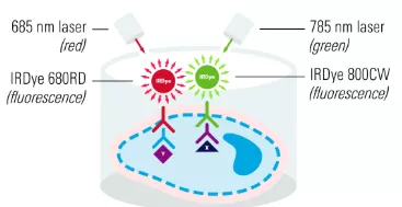
In-Cell/On-Cell Western
In-Cell/On-Cell Western的实验流程
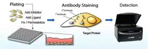 | |
实验流程步骤 | |
以一定密度在96/384孔板上培养细胞 | |
2 | 生长到合适密度后,加药物或其他处理 |
3 | 加入多聚甲醛进行固定 |
4 | 加入Triton X-100对细胞进行透化处理 |
5 | BSA封闭30 min |
6 | 加入一抗孵育1h |
7 | 加入荧光标记二抗,洗涤检测 |
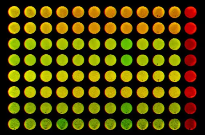
In-Cell/On-Cell Western提高高通量实验的可重复性和准确性
HeLa细胞分别用10uM的CAM和DMSO处理7天,In-Cell Western检测氧化磷酸化复合物的的水平,统计学数据表明,对照组、DMSO和CAM处理组的变异系数CV均较低,重复性好。
GAPDH和PMLC20通过WB分别在两张膜上各做13个重复,变异系数为0.27;GAPDH和PMLC20通过ICW检测,30个重复,变异系数为0.08-0.16。
与化学发光膜WB法相比,ICW的通量更高,变异系数更低,提高实验结果重复性和准确性。
与膜上WB法相比, In-Cell/On-Cell Western更快速便捷
In-Cell/On-Cell Western的应用举例
01 | Insulin Rearch |
02 | Signaling Pathways |
03 | Drug Rearch |
04 | RNAi |
05 | Apoptosis |
06 | Ion Channel |
01.Insulin Rearch
Dose-dependent results of human insulin receptor activation. A. Images of ICW signals detected by an Odyssey® Infrared Imaging System (LI-COR, Bad Homburg, Germany) with the 700- and 800-nm channels. The signals obtained from DNA and cell stain with DRAQ5™ and Sapphire700™ excited at ~680 nm and detected at ~700 nm are displayed in red, whereas the signals obtained from detecting the phosphorylated human insulin receptor (p-hIR) excited at ~780 nm and detected at ~800 nm are displayed in green. The composite overlay shows both measurements in one image.
值得注意的是,胰岛素筛选主要是基于胰岛素刺激细胞后,对细胞膜受体磷酸化水平进行检测,因其对细胞培养需求大,普通WB难以实现,而LI-COR Odyssey 的In-Cell Western能对人胰岛素和胰岛素类似物的效价进行高灵敏度的定量检测,因此美国药典(United States Pharmacopoeia,USP)推荐用LI-COR Odyssey的In-Cell Western方法进行胰岛素筛选。
02.Signaling Pathways
Effects of Y27632 and fasudil on p-ERK protein expression levels in cultured spinal cord microglia.(A) In-cell western blot assay of the changes in p-ERK protein expression levels in response to Y27632 (10 μM) and fasudil (41 μM) treatments at 1, 3 and 6 hours. (B) Statistical analysis of p-ERK protein levels. [1]
03.Drug Rearch
Tim-3 is mostly located inside resting THP-1 cells but is externalised after PMA-dependent PKCα activation. Tim-3 was measured using a Li-Cor on cell assay with (in cell western) or without (on cell assay) methanol permeabilisation (see the Materials and methods section). Exposure to the anti-Tim-3 antibody was performed for 2 h. Images are from one experiment representative of five which gave similar results.[2]
04.siRNA
Representative in-cell western dual detection of K18 protein (green), β-actin protein (red), and combined (merged image-yellow) following exposure to either Lipofectamine™ or equimolar concentrations of a non-targeting siRNA (siCTL) as controls, or 100 nM siRNA to KRT8/18 (siKRT8/18). Diminished expression of K18 in the single and merged images was evident following 72 h exposure to siKRT8/18.[3]
05.Apoptosis
Following the pre-incubation of HK-2 cells with 10, 20 and 40 µM SchB for 2 h, the cells were stimulated with cis-DDP for 24 h. The activation of caspase-3 was then determined by in-cell western blot analysis. [4]
06.Ion Channel
Detection of P‐Akt, the active (phosphorylated) form of Akt within TH+ neurons using infrared fluorescence imaging applied to the In‐Cell western blot technique in 7 DIV cultures exposed chronically to GLIB in the presence or not of LY applied only acutely (20 min). [5]
In-Cell/On-Cell Western 应用广泛
高通量快速的In-Cell/On-Cell Western已广泛应用到细胞自噬、凋亡、癌症、药物研究、信号转导、离子通道、基因表达等多个方面。
In-Cell/On-Cell Western应用文献还可登录LiCor官网查询
https://www.licor.com/bio/publications/
In-Cell/On-Cell Western的优势
01 | 高通量,一次可实现96/384样品检测 |
02 | 可用不同荧光标记二抗同时检测两个蛋白 |
03 | 可做大分子量细胞蛋白检测 |
04 | 特异性高、灵敏性好,大大节约实验时间 |
05 | 重复性好、精确度好 |
参考文献
1. The Rho-associated kinase inhibitors Y27632 and fasudil promote microglial migration in the spinal cord via the ERK signaling pathway[J]. Neural Regeneration Research, 2018, v.13(04):111-117.
2. Yasinska I M , Ceccone G , Ojea-Jimenez I , et al. Highly specific targeting of human acute myeloid leukaemia cells using pharmacologically active nanoconjugates[J]. Nanoscale, 2018, 10.
3. Trisdale S K , Schwab N M , Hou X , et al. Molecular manipulation of keratin 8/18 intermediate filaments: modulators of FAS-mediated death signaling in human ovarian granulosa tumor cells[J]. Journal of Ovarian Research, 2016, 9(1):8.
4. Liu Q , Song J , Li H , et al. Schizandrin B inhibits the cis DDP nduced apoptosis of HK2 cells by activating ERK/NF?κB signaling to regulate the expression of survivin[J]. International Journal of Molecular Medicine, 2018.
5. Toulorge D , Guerreiro S , Hirsch E C , et al. KATP channel blockade protects midbrain dopamine neurons by repressing a glia-to-neuron signaling cascade that ultimately disrupts mitochondrial calcium homeostasis[J]. Journal of Neurochemistry, 2010, 114(2):553-564.
基因有限公司作为LI-COR近红外成像解决方案的合作伙伴与中国区独家代理商,与LI-COR一起邀请您一同关注近红外荧光成像技术在Western Blot准确定量、磷酸化等蛋白修饰化研究、In-Cell Western、双色EMSA、蛋白芯片及光免疫疗法等方面的应用进展。欲进一步了解详细内容,请关注我们的官方微信“基因快讯”或联系您身边的基因有限公司工作人员。